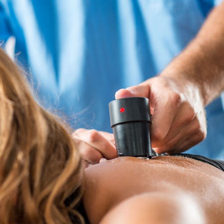
Ultrasound imaging (also known as diagnostic sonography or ultrasonography) is a medical diagnostic technique that involves the use of high-frequency sound waves to produce images of the internal structures (such as joints, muscles, ligaments, tendons, nerves, internal organs and soft tissues) within your body. It is used to help healthcare providers evaluate, diagnose and treat musculoskeletal (MSK) injuries that may or may not be visible on X-Rays such as sprains, strains and tears. Because ultrasound can also scan bone surfaces, it is used as an alternative to X-Rays to detect fractures of the wrist, elbow and shoulder in pediatric patients up to age 12.
Ultrasound images capture the structure and movement of the body's internal organs, as well as blood flowing through blood vessels and arteries. X-Rays and MRIs can't. An ultrasound image can follow a structure from its origin to where it attaches to another structure, so musculoskeletal ultrasounds are ideal for examining long structures, such as nerves and muscles. It does not use radiation, as X-Rays do, so it is safe for imaging areas where radiation sensitivity is a concern. It has been used for years in gynecology to examine female pelvic organs and in obstetrics to determine the gestational age and level of fetal development, confirm fetal viability, determine the presence or absence of birth defects and for other internal examinations concerning the pregnancy.
Ultrasound Imaging is used to diagnose and treat a variety of conditions including:
- Muscle, ligament and tendon tears
- Bursitis and tendinitis of the rotator cuff in the shoulder, Achilles tendon in the ankle and other tendons throughout the body
- Bursae and joint inflammation or fluid (effusions)
- Early changes of rheumatoid arthritis
- Nerve impingements/entrapments
- Knee conditions, such as Patellar Tendonitis, ACL tear, Meniscal tear, IT Band Syndrome, Popliteal Tendinitis and Baker's Cyst
- Foot conditions, such as Plantar Fasciitis, Metatarsalgia and tendon/ligament sprains and strains
- Cartilage defects in the knee at the femoral condyle and meniscus
- Cartilage defects in the labrum of the shoulder and hip
Musculoskeletal ultrasound imaging can complement MRI imaging and sometimes even eliminate the need for unnecessary and expensive MRIs. Using ultrasound in place of MRI does not compromise diagnostic accuracy and can reduce healthcare costs significantly.
For more information on Musculoskeletal Ultrasound imaging or to set up an appointment, call us at (630) 686-1000 or request an appointment with us online.
